

Protein Research Group of the Laboratory of New Methods for Biology of the IBI RAS invites graduate students (master’s degree) to join postgraduate course in molecular biophysics.
KM-7 micromanipulators are designed for providing very accurate moving of microtools in the field of view of a microscope in order to operate on microobjects. They comprise mechanical and air-operated micromanipulators differing by their principle of transmitting hand movements to microtools.
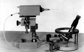
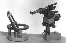
The micromanipulators are placed next to a microscope on an antivibration base plate and locked on it with the help of their magnetic base. According to a customer's order, the micromanipulators can be adapted to an inverted microscope.
The set of micromanipulators and microinjectors is highly accurate equipment for embryogenetic investigations and technologies as well as for practical work in the field of artificial impregnation and cloning.
Mechanical micromanipulators provide
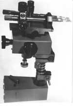
They are in a wide use in microelectrode investigations.
They are in a wide use in cellular microsurgery.
They provide dividing a cell into two parts with the use of a caprone microligature of 5 to 20 μm in diameter.
Air-operated and piston devices for microinjections are designed for exhaustion and injection of micro-volumes of liquid.
The set of microinjectors and micromanipulators is highly accurate equipment for embryogenetic investigations and technologies as well as for practical work in the field of artificial impregnation and cloning.

It can be placed on any micromanipulator and is ideal for injection of micro-volumes of liquid of 10-8 to 10-12 ml with the help of compressed air. Remote control from a control unit.
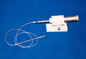
It can be placed on any micromanipulator and allows a exhaustion and injection of micro-volumes of liquid of 10-5 to 10-8 ml. The remote control is performed with a pneumatically driven knob.

An instrument for vitrification of embryos and oocytes is designed for transformation of liquid phase of biological objects (cells, tissues) into a solid phase without formation of ice crystals in intercellular and intracellular space.
Biological objects pretreated by cryoprotectors are placed into liquid nitrogen with the temperature less than -196ºC. The volume above liquid nitrogen in hermetically closed thermostat is vacuumized, which results in a decrease in temperature down to about -210ºC. The vitrification simplifies the process of cooling and excudes the risk of physical and chemical damages because of water crystallization. Pregnancy rate after vitrification is about 16%.
| Volume of liquid nitrogen chamber: | 500±50 cm³ |
| Liquid nitrogen temperature: | -196 ºС to -206 ºС ±4 ºС |
| Overall size of cryogenic module: | 350×350×310 mm |
| Overall size of vacuum pump: | 250×216×165 mm |
| Macc of of cryogenic module: | 6.5 kg |
| Mass of vacuum pump: | 9.0 kg |
| Power: | 250 W |

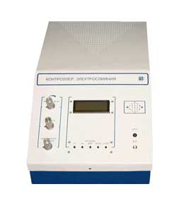
The instrument is designed for generation of cells and embryos with changed genetical properties by means of electroporative fusion. The fusion of cells through pores occures under strong electric field. The instrument has two channels of generation of high voltage impulses from 550 V to 3000 V with separate output connectors. The instrument control is achieved by a microprocessor and the output parameters are shown on LCD.
The instrument is composed of an electroporation controller and a chamber for cell fusion.
| Amplitude of output impulses: | I range 100-500 V; II range 500-3000 V |
| Pulse duration: | I range 300 μs - 99 ms; II range 5-99 ms |
| Number of impules in a series: | 1 to 99 |
| The chamber: | The chamber is placed in a standard 90 mm Petri dish. The distance between electrodes can be changed with a step of 2 mm |
| Power supply: | 220 V, 50 Hz |
| Power: | 200 W |
The instrument is designed for generation of cells and embryos with changed genetic properties by means of electrical breakdown in membrane. The instrument is composed of a chamber for electrostimulated cell fusion and a controller of the electrofusion.
The efficiency of electrofusion is achieved due to tight pressing of wire electrodes to a glass slide and the ability to tighten them by special tap wrenches. The spacing between the wire electrodes is from 50 to 500 μm. The elctrodes are made of platinum- iridium or nichrome wire with 0.2 to 0.3 mm in diameter. Chamber material is biologically inert and allows cold sterilization by alcohol-containing means. The chamber can be placed on the table of a microscope for visual control of the fusion process.
The fusion controller generate electric signals needed for the fusion process. Harmonic wave with frequency 250 kHz to 2.5 MHz and amplitude 20 V is used for cell aligning. The duration of the aligning signal can be from 1 to 9999 s. Before the end of the process the signal amplitude increases by 10% to press cells to each other tightly. A signal with amplidude from 50 to 300 V and duration from 1 to 9999 μs (number of impulses from 1 to 9) is used for electrical breakdown in the membrane. After the breakdown the electrofusion controller generates a harmonic damped wave with the frequency and amplitude of the aligning signal, which decays to zero during the action of the first signal. This mode assures the best conditions for survival of the reconstructed cell. The electrofusion controller is conrolled by a microprocessor.
Overall dimensions of the electrofusion controller are 245×435×165 mm.

The instrument for cryoconservation is designed for superfast freezing of droplets of suspension of animal or plant cells and for collection of the freezed droplets to a container for a long storage at extremaly low temperatures. The instrument is mounted on a neck of a Dewar flask.
The device can be successfully used for creation of cryo-banks in scientific laboratories, natural reserves, and farms.
| Volume of a vessel for cell suspensions and of a comtainer for collection of frozen droplets: |
up to 100 mL |
| Freezing temperature: | up to -70ºC |
| Rate of droplets formation: | 0.5 to 2 droplets/s |
| Source of freezing: | Dewar vessel with liquid nitrogen |
| Overal dimensions: | 450х250х400 mm |
Protein Research Group of the Laboratory of New Methods for Biology of the IBI RAS invites graduate students (master’s degree) to join postgraduate course in molecular biophysics.
A conference of young scientists and students "Biomedical engineering – 2010" will take place in IBI RAS in November 2010.